Tampilkan postingan dengan label diabetes. Tampilkan semua postingan

Rabu, 22 Juni 2016

Can Stevia Help You Ward Off Type II Diabetes? A Review

Unfortunately, it is not even clear if you need the "white stuff", i.e. pure steviosides, whole leaves of leaf-extracts to maximize the anti-diabetic effects of stevia. What is clear, though, is that there's still a lot of research to be done.
"Can Stevia Help You Ward Off Type II Diabetes?" That's not just the title of today's SuppVersity article, it is also the research question of a recent paper by Esteves A.F. dos Santos from Farmácia Progresso (dos Santos. 2016). An interesting question with an obvious answer: if you replace sugar in your diet with stevia, it will help.

Now, you know that this would not be worth discussing in a SuppVersity article of its own. What is worth discussing, though, is that stevia contains "compounds and other substance obtained from stevioside hydrolyses" (dos Santos. 2016) such as isoteviol of which studies show that they can be used as 'active' diabetes treatments - meaning: they help, even if you take them on top of sugar / your regular diet.
You can learn more about sweeteners at the SuppVersity

Aspartame & Your Microbiome - Not a Problem?

Will Artificial Sweeteners Spike Insulin?

Sweeteners & the Gut Microbiome Each is Diff.

Chronic Sweeten-er Intake Won't Effect Microbiome

Stevia, the Healthy Sweetener?

Sweeteners In- crease Sweet- ness Threshold
To understand how stevia may help you to ward off diabetes, you will first have to understand how the latter actually develops. In the previously references review, dos Santos writes about the consequences of life-style induced weight gain and concomitant increases in body fat and insulin resistance (IR):
Figure 1: Illustration of the etiology of type II diabetes from a secondary source (in dos Santos. 2016)
"After an initial increase of insulin production as a response to IR in peripheral tissue, pancreatic β cells no longer have the ability to control glucose homeostasis, leading to endocrine sys-tem imbalances. Under glucagon influ-ence, the liver contributes significantly in glucose homeostasis because liver makes the balance between capture / storage of glucose, via glycogenesis, and the release of glucose by glycogenolysis and gluconeogenesis. 
Constant, prolonged state of hypergly-cemia enables the formation of Advanced Glycation End-Products (AGEs). AGEs are responsible for the onset of diabetic complications, such as neurological and kidney complications (diabetic nephropathy), aging and cardiovascular complications: dyslipidemia, hyperten-sion, [etc.]" (dos Santos. 2016).
The previously referenced AGEs and the significant increase of reactive oxygen specimen (ROS), which leads to decreased levels of antioxidants enzymes, increase lipid peroxidation, will increase the risk for cardiovascular diseases and exacerbate the state of the disease, which has - at this point - started to self-propel its own progression.

If using stevia could break this vicious cycle, this would obviously be awesome!

Initial evidence that suggests that stevia could do what the subheading suggests, and decrease blood glucose levels comes from ... you guessed it: rodents. In a 4 week supplementation study, rats who were fed Stevia rebaudiana extract - combined with high-carbohydrate and high-fat diets - exhibited a sign. lower increase in glucose and worsening of their glucose tolerance in an oral glucose tolerance test (OGTT) - a result that was soon confirmed in human beings who ingested an infusion of 5 Stevia rebaudiana leaves for 3 days, every 6 hours (see Figure 2):
Figure 2: Effect of stevia leaf extract (5g) blood glucose of 16 healthy subjects on oral glucose tolerance test (Curi. 1986).
Similar results have been observed by Anton et al. (2010) who compared the effect of preloads of stevia with preloads of other sweeteners, such as aspartame or sucrose in obese and normal subjects. As the data in Figure 3 shows, these preloads, which were consumed by study participants 20 minutes before their test lunch and dinner meals, decreased postprandial insulin significantly.
Figure 3: Blood glucose response in man with preloads of either sucrose, aspartame or stevia (Anton. 2010)
Now, the obvious question we have to answer is: how did that work? There are different speculative and proven mechanisms that could contribute to the anti-diabetic effects of stevia:

  • one study showed that Stevia rebaudiana will inhibit the pancreatic enzyme alpha-amylase and alpha-glucosidase and thus the breakdown of carbs in the intestine (Adisakwattana. 2010),
  • Figure 4: Effects of Stevia extracts on glucose transport activity compared to the effect of insulin. SH-SY5Y (a) and HL-60 (b) cells were treated with steviol glycosides (1 mg/mL), with 100 nM insulin (I), with steviol glycosides and insulin simultaneously, or 1 mM standard compounds (StReb, StStev | Rizzo. 2013).
    stevia rebaudiana extracts may also act similar to insulin and are equally effective in increasing glucose uptake,because the co-treatment with insulin and stevia extracts increase glucose uptake significantly higher than the increase due to insulin alone (Rizzo. 2013), , similar results were reported by Akbarzadeh et al. (2015) in STZ-induced diabetic rats
  • various studies provide evidence for the anti-oxidant effects of stevia and respective extracts, which will - in view of the inflammatory nature of type II diabetes - obviously contribute to its anti-diabetic effects
  • at least one study shows that isostevial, one of the stevia glycosides, appears to work part of its magic via activating the PPAR receptor alpha (Xu. 2012)
Whether there is one specific agent that is responsible for the previously listed effects is still debated. Among the "suspects" are primarily steviol glycosides for which anti-hyperglycemic effect has been observed in doses ranging from 5 mg / kg to 200mg/kg (González. 2014)
Is stevia even safe? You will be surprised to hear that, but the safety of the chronic consumption of stevia, the "natural sweetener", cannot be guaranteed (see possible ill effects on fertility). While studies in adult hypertensive patients show that it is "likely safe" when taken orally (250-500mg stevioside) thrice daily for up to two years, scientists argue that it could be "possibly unsafe, [...w]hen taken [by] children, or pregnant or lactating women or for periods longer than two years, due to insufficient available evidence" (Ulbricht. 2010). The same goes for its use by patients with hypotension, hypocalcemia, hypoglycemia, or impaired kidney function. In view of what we know about the possibility of allergy/hypersensitivity to other members the daisy family (Asteraceae/ Compositae), one may also suspect that allergic reactions, which have not been reported in the literature, yet, are not likely.
More specifically, these compounds have been observed to offset "the glucagon hypersecretion by pancreas α cells that's usually caused by prolonged exposure to fatty acids, and changed genes expression responsible for the metabolism of fatty acids" (dos Santos. 2016). They have also been shown to increase the glucose uptake of pancreatic cells, thus rendering them more sensitive to (small) changes in blood glucose levels; and Gonzalez et al. found them to be capable of increasing proinsulin mRNA concentration and insulin in pancreas INS-1 cells - with the result being a sign. increase the content of insulin in cells.
Figure 5: Glucose (left) and lipid (right) levels in rodents after 14 days on a high fat diet w/ different amounts of isosteviol in the diet - the effects are sign., but the effect size is small (Xu. 2012)
Of the various steviosides, dos Santos highlights isosteviol, a stevioside hydrolyzate, in particular, because it has been shown to have especially pronounced influence on glucose metabolism (Xu. 2012) in a 14-day rodent study in which the animals were fed high-fat chow and the oral administration of  isosteviol orally administrated at doses from 1 to 5 mg/kg/day led to a statistically significant decrease in insulin levels, accelerated glucose clearance and improved insulin sensitivity while simultaneously lowering total and LDL cholesterol and increasing HDL - not bad even if the effect size is relatively small, right?
"The mechanism underlying these effects may be related to the expression of PPARα, since this has changed in the treatment with isosteviol. Furthermore, the pretreatment with isoteviol improves antiapoptosis factor Bcl-2 expression and inhibits the NF-kB expression, and increases SOD and GSH-PX activity. Isosteviol has anti-inflammatory effects, which may possibly be related to hypoglycemic effect and the ability to change lipid profile" (dos Santos. 2016).
Unfortunately, the results Xu et al. presented 4 years ago still await confirmation in human studies. The same goes for the first stevia based anti-diabetes "drugs" which seek to increase the bioavailability (in serum) of steviosides by bioconjugating them on biodegradable Pluronic-F-68 copolymer based PLA nanoparticles by the means of nanoprecipitation (Barwal. 2013). These studies exist, like a recent study by Kassi et al. who introduced low glycemic load snacks based on Stevia to a low calorie diet in patients with metabolic syndrome and found this to be a safe and highly efficient means to "further reduc[e] BP [blood pressure], fasting glucose, ox[idized] LDL and leptin compared to a hypocaloric diet alone, decreasing, thus, further the risk of atherosclerosis and DMT2" (Kassi. 2016) - as part of a regular diet and in place of high sugar foods, stevia is thus the most effective.
Figure 6: One of the few long(er) term studies in (diabetic) humans found no effect of 1g rebaudioside on glycemia (Maki. 2008) - so, don't get too excited about stevia being the new metformin.
So what's the verdict then? Well, I guess you won't be happy if I say that more research is, as usually, necessary. Dos Santos is yet right that "Stevia rebaudiana is a good option to be included in the group of nutraceuticals", in view of its "action and its main compounds (stevioside and rebaudioside A) concerning glycaemia control, diabetes consequences, and early development of IR" (dos Sanots. 2016).

In as much as it can be considered a "medicinal herb," though, its safety of and necessity of higher dosages, as well as the exact mechanism of action require further investigation. Whether it makes sense to develop sustained released, high bioavailability 'stevia drugs' does yet appear questionable to me. - in particularly, because isosteviol "is not subject to intestinal hydrolysis and has shown results as therapeutic agent for type 2 diabetes and its consequences" (dos Santos. 2016), without being chemically / molecularly altered - using "regular" stevia and that to replace sugar does therefore still appear to be the best 'anti-diabetic' use for this sweetener which is up to 150 times sweeter than sugar, heat- and pH-stable, and not fermentable | Comment on Facebook!
References:
  • Adisakwattana, Sirichai, et al. "Evaluation of α-glucosidase, α-amylase and protein glycation inhibitory activities of edible plants." International Journal of Food Sciences and Nutrition 61.3 (2010): 295-305.
  • Akbarzadeh, Samad, et al. "The Effect of Stevia Rebaudiana on Serum Omentin and Visfatin Level in STZ-Induced Diabetic Rats." Journal of dietary supplements 12.1 (2015): 11-22.
  • Anton, Stephen D., et al. "Effects of stevia, aspartame, and sucrose on food intake, satiety, and postprandial glucose and insulin levels." Appetite 55.1 (2010): 37-43.
  • Barwal, Indu, et al. "Development of stevioside Pluronic-F-68 copolymer based PLA-nanoparticles as an antidiabetic nanomedicine." Colloids and Surfaces B: Biointerfaces 101 (2013): 510-516.
  • Curi, R., et al. "Effect Of Stev/A Reba Ud/Ana On Glucose Tolerance. In Normal Adult Humans." Braz. j. med. biol. res (1986).
  • González, et al. "Stevia rebaudiana Bertoni: a potencial adjuvant in the treatment of diabetes mellitus." CyTa – Journal of Food 12.3 (2014): 218- 226.
  • Kassi, Eva, et al. "Long-term effects of Stevia rebaduiana on glucose and lipid profile, adipocytokines, markers of inflammation and oxidation status in patients with metabolic syndrome." (2016).
  • Maki, K. C., et al. "Chronic consumption of rebaudioside A, a steviol glycoside, in men and women with type 2 diabetes mellitus." Food and Chemical Toxicology 46.7 (2008): S47-S53.
  • Rizzo, Benedetta, et al. "Steviol glycosides modulate glucose transport in different cell types." Oxidative medicine and cellular longevity 2013 (2013).
  • Ulbricht, Catherine, et al. "An evidence-based systematic review of stevia by the Natural Standard Research Collaboration." Cardiovascular & Hematological Agents in Medicinal Chemistry (Formerly Current Medicinal Chemistry-Cardiovascular & Hematological Agents) 8.2 (2010): 113-127.

Rabu, 18 Mei 2016

Can Oxidized Proteins Kill You? PROTOX Links Processed High Protein Foods to IBS, Diabetes, Cancer, NAFLD & Co.

Dietary protein sources: You better eat them before they're rancid.
There's such a thing as "protein oxidation"? If you are asking yourself this question, you will probably have missed the 20th century studies by Henry D. Dakin (*1880–†1952). Dakin originally reported the oxidative degradation of particular amino acids during digestion and introduced the potential biological consequences of such biochemical reactions.

The impact of PROTOX, as this form oxidation is called to distinguish it from the way better known LOX (lipid oxidation) on human health was, at that moment, wholly unknown.

As Estévez and Luna point out in a recent paper in the peer-reviewed scientific journal "Critical Reviews in Food Science and Nutrition", PROTOX has been in the focus during the succeeding decades, though, "owing to the association between the oxidative damage to proteins and aging and age-related diseases (Berlett & Stadtman, 1997)" (Estévez. 2016).
Learn more about meat at the SuppVersity

You May Eat Pork, too!

You Eat What You Feed!

Meat & Prostate Cancer?

Meat - Is cooking the problem

Meat Packaging = Problem?

Grass-Fed Pork? Is it Worth it?
Earl R. Stadtman (*1919–†2008), a renowned biochemist of the 20th century and mentor of various Novel-prized scientists, was one of the pioneers in unveiling the chemistry and biological consequences of PROTOX. From the elucidation of mechanisms whereby the rates of metabolic reactions match to the necessities of the living cell, he identified the connection between unbalanced oxidative metabolism (≈ oxidative stress) and impaired physiological processes (Stadtman, 1990).
Figure 1: Oxidative damage to poultry: Sources of oxidative stress, impact of oxidation, and antioxidant strategies (Estévez. 2015).
"While some of the underlying mechanisms of the connection between in vivo PROTOX and disease are still to be clarified, it is accepted that PROTOX plays a role in aging and age related diseases such as Alzheimer’s, Parkinson’s, inflammatory Bowel’s (IBD), rheumatoid arthritis, diabetes, muscular dystrophy, and cataractogenesis, among others (Berlett & Stadtman, 1997). 
On account of the effort of brilliant scientists, the ‘poor cousin’ of lipid oxidation is now extolled as a topic of the utmost scientific interest" (Estévez. 2016).
Now that you know all that, I suspect that you are asking yourself what this "protein oxidation" has to do with "Food Science and Nutrition". Well, the answer is actually pretty simple: While PROTOX has been for decades disregarded as a major cause of food deterioration, it does play a major role in foods from nutritional, sensory and technological points of view.
Note: There will be a follow up to this article, next week with answers to your questions, such as (1) How can I avoid protein oxidation when preparing protein containing meals? (2) Which foods are the most susceptible? (3) If processing is an issue won't protein powders be the worst offenders? Not your question? Feel free to post additional questions you may have here.
In the early years of the 21st century, numerous subsequent studies shed light on the oxidative modifications undergone by muscle proteins during handling, processing and storage of muscle foods; and among of the better known results of these studies are...
Figure 2: Hypothesis of the influence of dietary protein oxidation on in vivo oxidative stress and pathological conditions. "It is actually well-established that the composition of food and the dietary habits have physiopathological consequences" (Estévez. 2016).
  • that the formation of PROTOX will impair the functionality and digestibility of meat and dairy proteins (Santé-Lhoutellier et al., 2007; Feng et al., 2015),
  • that the presence of PROTOX will impair the nutritional value and sensory attributes of muscle foods such as tenderness (Bao & Ertbjerg, 2015) and flavor (Villaverde et al., 2014), and the chemistry behind food PROTOX, the occurrence and consequences of PROTOX during food
  • that PROTOX will almost inevitably occur during storage and processing, but can be reduced by applying certain strategies (Bekhit et al., 2013; Estévez, 2015; Soladoye et al., 2015).
As Estévez and Luna point out, the investigation of postprandial events, which has started, only recently, "enables a more realistic approach to investigate the impact of food intake on nutrition and health as food components are severely modified during the digestion phases" (Estévez. 2016). Unfortunately, many of the currently existing studies investigated events in-vitro. The important (and certainly most relevant) question, whether the consumption of oxidized proteins from food can actually harm you, however, has been addressed by a limited number of studies, only.
How to prevent protein oxidation? There's no way you prevent the oxidation of proteins in food completely, but packaging in light-blocking low-oxygen containers and not processing foods like crazy (exerting force on it in a grinder, for example | see Figure, right. Bao. 2015) could reduce the oxidation process just as significantly as not heating / burning meat will (Villaverde. 2013).
There's, nevertheless, "evidence that in vivo oxidation is a source of aging and disease calls to elucidate to which extent dietary oxidative stress contributes to aggravating in vivo oxidative stress and its harmful consequences" (Estéves. 2015); and these "harmful consequences" have been traced back to particular dietary oxidation products, of which researchers believe that they are able to induce or contribute to some pathological process in targeted cells or tissues through the induction of specific molecular responses (i.e. gene expression regulation).
  • Figure 2: Proposed mechanisms of pathogenesis exerted by dietary protein oxidation products. It was not until recently that the fact that dietary oxidized proteins would, themselves, be active executors of specific pathological processes was discovered (Estévez. 2016).
    the intake of foods high in PROTOX products, raises general oxidation markers, leads to cell damage and increases the risk of suffering health disorders such as coronary-heart diseases, neurodegenerative disorders and certain types of cancer (Esterbauer et al., 1992, 1993; Sies et al., 2005; Awada et al., 2012), 
  • interestingly, these processes have been linked to LOX products, as well, which turn out to be cross-linked to the cytotoxicity and mutagenicity potential of PROTOX species on the gastrointestinal tract or in internal organs upon absorption (Esterbauer et al., 1993), 
  • proteins are regarded as targets for post-translational changes, unlike LOX of which we believe that they have a direct damaging effect
  • the molecular basis of these processes commonly involves the interaction of primary and secondary LOX products (i.e. alkyl radicals, peroxides, hexanal, 4-HNE, MDA) with proteins of biological significance (formation of adducts) and other biomolecules such as DNA (Esterbauer et al., 1991; Awada et al., 2012).
  • cellular responses to these molecular changes usually imply the activation of particular signalling pathways that involves gene expression and/or suppression (Figure 2), 
Now, while all of this has been known for years, it was not until recently that the fact that dietary oxidized proteins and PROTOX products would, themselves, be active executors of specific pathological processes was discovered.
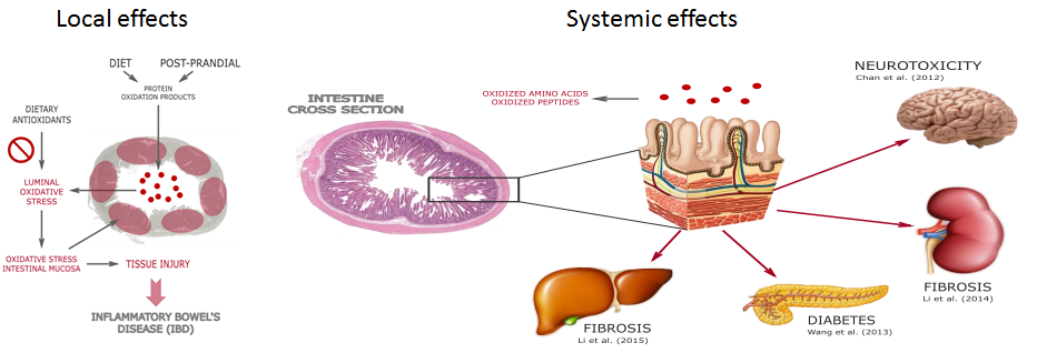
"The oxidation of food proteins during processing and storage leads to the inexorable accumulation of oxidation products that will be primary exposed to the gastrointestinal tract. As aforementioned, food PROTOX also occurs during consumption and gastrointestinal digestion increasing the concentration of oxidation products in the lumen. Scientific evidences support the impact of dietary oxidized proteins on intestinal flora disturbance, the redox state of intestinal tissues and the onset of local pathological conditions (Keshavarzian et al., 2003; Fang et al., 2012; Xie et al., 2014). 
Pierre et al. (2004), among others, already provided reasonable arguments to support the impact of luminal oxidative stress on cytotoxicity, genotoxicity and apoptosis in cells from colonic mucosa. More specifically, oxidative stress has been found to play a relevant role in the onset of carcinogenic processes, including CRC (Polyak et al., 1997; Valko et al., 2006). Interestingly, some clinical studies emphasize the extent of plasma protein carbonylation as a reliable marker of the risk of suffering CRC (Yeh et al., 2010; Chang et al., 2008). Chang et al. (2008) in particular, found altered protein carbonyl levels in CRC patients while LOX products remained at low levels. Others implicate the oxidative damage to proteins in the pathogenesis of CRC. This is the case of Nedic et al. (2013) who indicated the potential role of the carbonylation of insulin-like growth factor-binding proteins in CRC growth" (Estévez. 2016).
While the formerly cited evidence is mostly from in vitro studies, more recent data from rodents shows that intraperitoneal administration (= injection that is equivalent to oral consumption) of oxidized proteins to rats raised the level of advanced oxidation protein products )AOPPs) in the local intestine tissue and in blood inducing intestine epithelial death through a redox-dependent
pathway. As Estévez and Luna rightly point out, "[t]hese results proven that PROTOX products may be implicated in the transfer of oxidative stress from the luminal phase to the lamina propia of the intestinal mucosa facilitating the process of IBD" (Estézes. 2016 |see Figure 4, left).
Figure 4: LEFT - Pathogenesis of dietary protein oxidation products in the GIT: transfer of oxidative stress from lumen to intestinal mucosa, tissue injury and inflammatory disease.f, RIGHT - Absorption and subsequent pathological effects of dietary protein oxidation products in targeted tissues (Estévez. 2016).
The molecular mechanisms of this pathological effect involved is, according to the authors of this most recent review a NADPH oxidase-mediated ROS generation, JNK phosphorylation, and poly (ADP-ribose) polymerase-1 (PARP-1) activation. Consequences of which studies show that their effects are not limited to the gut.
Protein oxidation during refrigerated storage of liver pâtés with added BHT sage or rosemary essential oils (p < 0.05, between antioxidant groups within a day of storage denoted by letters | Estévez. 2006)
Vitamin E doesn't work, vitamin C only increases the formation of PROTOX! Studies suggest that adding known anti-oxidant to your foods may both promote and inhibit the formation of PROTOX or Pox, as they are also called. The usual suspects, such as tocopherols, however, will be failing you, here. Some phenolic-rich plant and fruit extracts have been shown to exert anti-oxidative protection of proteins in cooked pork patties, porcine liver pâté (see figure on the left) and chicken, but the pro-/anti-oxidative effect depends on the structure and the concentration of the respective phenolic compound.

In beef patties, a rosemary extract was found to have no protective effect against Pox and a mixture of ascorbate and citrate promoted Pox, while both anti-oxidant systems protected lipids from oxidation. Furthermore, addition of rosemary oil to frankfurters has been shown to inhibit Pox while addition of higher levels of the rosemary oil resulted in a prooxidative effect when the frankfurters were prepared with meat from white pigs showing that the anti-oxidative effect was dependent on concentration and product characteristics. Lastly, it should be mentioned that the synthetic hydrophilic anti-oxidant Trolox (a vitamin E analogue) was found to prevent oxidation of both protein and lipid fractions (Lund. 2011).
As such, diets rich in readily oxidized components (polyunsaturared fatty acids) and meat proteins are believed have long been linked to a increased risk of suffering various forms of IBD such as Crohn's disease and ulcerative colitis (Hou et al., 2011), but it is also, as Estévez and Luna point out, also reasonable to hypothesize that such diets may contribute considerable oxidized proteins given the close association between LOX and PROTOX in food systems and in the gastrointestinal tract (Soladoye et al., 2015; Van-Hecke et al., 2015).
"Gurer-Orhan et al. (2006) already hypothesized that oxidized amino acids may be misincorporated into proteins such as enzymes and structural element in cells, potentially contributing to malfunction, cell apoptosis and disease. These authors emphasized that post-translational oxidative modification of proteins may not be the only factor that contributes to in vivo PROTOX suggesting that external (dietary) sources of oxidized amino acids may cause direct toxic effects by being used for de novo synthesis of proteins. To similar conclusions came succeeding studies carried out by Dunlop et al. (2008; 2011). The absorption and subsequent deleterious effects of unnatural oxidized amino acids such as meta-tyrosine and 3,4-dihydroxyphenylalanine (L-DOPA) are known to occur in animals and humans leading to dysfunctional proteins and toxicity (Dunlop et al., 2015). These species may not only be formed in foods as a result of tyrosine oxidation, they are also natural components of edible plants and beans (Siddhuraju & Becker, 2001; Davies, 2003; Dunlop et al., 2015). Chan et al. (2012) demonstrated that substitution of L-tyrosine residues in proteins with L-DOPA causes protein misfolding, promotes protein aggregation and stimulates the formation of autophagic vacuoles in SH-SY5Y neuroblastoma cells. Other oxidized forms of tyrosine, such as the ortho-tyrosine, contribute to the impairment of the insulin-induced arterial relaxation through the attenuation of endothelial nitric oxide synthase (eNOS) phosphorylation (Szijártó et al., 2014)" (Estévez. 2016).
Similar effects as they are described for oxidized tyrosine have been observed for oxidized tryptophan and lysine, which are present in significant amount in a plethora of processed foods including, but not restricted to meat and dairy.
Table 1: Relation of 2-AAA (oxidized lysine) levels to the risk of future diabetes in the whole sample and subgroups of 188 individuals who developed diabetes and 188 propensity-matched controls from 2,422 normoglycemic participants followed for 12 years in the Framingham Heart Study (Wang. 2013).
With respect to the latter, i.e. oxidized lysine, it is certainly worth poining out that 12-years long metabolomic study with human patients found this compound to be the most reliable indicator of diabetes risk - plus: Wang et al. were able to demonstrate that its oral ingestion increased the levels of the oxidized amino acid particularly in the pancreas, the same organ that is failing in diabetes (Wang et al., 2013). Other oxidized amino acids have been linked to
  • cell death in the intestine, colon and small intestine and subsequent irritable bowel disease (various) AOPPs; Xie et al (2014), Fang et al. (2012), Keshavarzian et al. (2003), Wu et al. (2015)
  • intestinal flora & redox state disturbance and liver & kidney stress, oxidized casein; Fang et al. (2012), Li et al. (2013), and Li et al. (2014)
With oxidized proteins and amino acids, there's thus yet another, often overlooked parameter of our food intake and dietary habits with "straightforward impact on health status" (Estevéz. 2016).
Bottom line: As premature as our understanding of the biology that governs the beneficial/detrimental effects of certain dietary components still is, there is ample evidence that not just the consumption of oxidized dietary fats, but also that of proteins, the major components of most foods (particularly animal-source), could harm us.

Fresh (!) Red Meat Acquitted - Overgeneralized Accusations that Red Meat Consumption Triggers Cancer Overlooks Influence of Processing & Other Confounding Factors | more
This does not mean that proteins (animal or plant proteins) are still a vital part of a healthy diet - that's indisputable. As Estevez and Luna point, out, "the discussion about dietary proteins [wich] is typically centered in the quantity, quality (≈ amino acid profile; biological value) and bioavailability upon digestibility," future recommendations for protein intake will have to consider the differential impact of foods with different PROTOX levels, as well. In this context, it should be obvious why "the current increase of the intake by population of highly processed animal-based foods with high protein content and presumably high oxidation rates" has been found to predict the raise of health disorders already associated to in vivo or dietary oxidative stress, in dozens if not hundreds of epidemiological studies | Comment!
References:
  • Awada, Manar, et al. "Dietary oxidized n-3 PUFA induce oxidative stress and inflammation: role of intestinal absorption of 4-HHE and reactivity in intestinal cells." Journal of lipid research 53.10 (2012): 2069-2080.
  • Bao, Yulong, and Per Ertbjerg. "Relationship between oxygen concentration, shear force and protein oxidation in modified atmosphere packaged pork." Meat science 110 (2015): 174-179.
  • Berlett, Barbara S., and Earl R. Stadtman. "Protein oxidation in aging, disease, and oxidative stress." Journal of Biological Chemistry 272.33 (1997): 20313-20316.
  • Chan, Sandra W., et al. "L-DOPA is incorporated into brain proteins of patients treated for Parkinson's disease, inducing toxicity in human neuroblastoma cells in vitro." Experimental neurology 238.1 (2012): 29-37.
  • Chang, Dong, et al. "Evaluation of oxidative stress in colorectal cancer patients." Biomedical and Environmental Sciences 21.4 (2008): 286-289.
  • Davies, Michael J. "Singlet oxygen-mediated damage to proteins and its consequences." Biochemical and biophysical research communications 305.3 (2003): 761-770.
  • Davies, Michael J. "The oxidative environment and protein damage." Biochimica et Biophysica Acta (BBA)-Proteins and Proteomics 1703.2 (2005): 93-109.
  • Dunlop, Rachael A., Roger T. Dean, and Kenneth J. Rodgers. "The impact of specific oxidized amino acids on protein turnover in J774 cells." Biochemical Journal 410.1 (2008): 131-140.
  • Dunlop, Rachael A., Ulf T. Brunk, and Kenneth J. Rodgers. "Proteins containing oxidized amino acids induce apoptosis in human monocytes." Biochemical Journal 435.1 (2011): 207-216.
  • Estévez, Mario, Sonia Ventanas, and Ramón Cava. "Effect of natural and synthetic antioxidants on protein oxidation and colour and texture changes in refrigerated stored porcine liver pâté." Meat science 74.2 (2006): 396-403.
  • Estévez, M. "Oxidative damage to poultry: from farm to fork." Poultry science 94.6 (2015): 1368-1378.
  • Estévez, M., and C. Luna. "Dietary Protein Oxidation: A Silent Threat to Human Health?." Critical Reviews in Food Science and Nutrition just-accepted (2016): 00-00.
  • Esterbauer, Hermann, et al. "The role of lipid peroxidation and antioxidants in oxidative modification of LDL." Free Radical Biology and Medicine 13.4 (1992): 341-390.
  • Esterbauer, Hermann. "Cytotoxicity and genotoxicity of lipid-oxidation products." The American journal of clinical nutrition 57.5 (1993): 779S-785S.
  • Fang, W., et al. "Effect of oxidated food protein on mice gut floraand redox state." Chinese Journal of Microecology 24 (2012): 193-196.
  • Gurer-Orhan, Hande, et al. "Misincorporation of free m-tyrosine into cellular proteins: a potential cytotoxic mechanism for oxidized amino acids." Biochemical Journal 395.2 (2006): 277-284.
  • Hou, Jason K., Bincy Abraham, and Hashem El-Serag. "Dietary intake and risk of developing inflammatory bowel disease: a systematic review of the literature." The American journal of gastroenterology 106.4 (2011): 563-573.
  • Keshavarzian, A., et al. "Role of reactive oxygen metabolites in experimental colitis." Gut 31.7 (1990): 786-790.
  • Keshavarzian, A., et al. "Increases in free radicals and cytoskeletal protein oxidation and nitration in the colon of patients with inflammatory bowel disease." Gut 52.5 (2003): 720-728.
  • Li, Zhuqing Leslie, et al. "Effect of oxidized casein on the oxidative damage of blood and digestive organs in mice." Acta Nutrimenta Sinica 35.1 (2013): 39-43.
  • Li, Zhuqing Leslie, et al. "Oxidized casein impairs antioxidant defense system and induces hepatic and renal injury in mice." Food and Chemical Toxicology 64 (2014): 86-93.
  • Li, Zhuqing Leslie, et al. "24-Week Exposure to Oxidized Tyrosine Induces Hepatic Fibrosis Involving Activation of the MAPK/TGF-β1 Signaling Pathway in Sprague-Dawley Rats Model." Oxidative medicine and cellular longevity 2016 (2015).
  • Lund, Marianne N., et al. "Protein oxidation in muscle foods: A review." Molecular nutrition & food research 55.1 (2011): 83-95.
  • Sante-Lhoutellier, Veronique, Laurent Aubry, and Philippe Gatellier. "Effect of oxidation on in vitro digestibility of skeletal muscle myofibrillar proteins." Journal of Agricultural and Food Chemistry 55.13 (2007): 5343-5348.
  • Siddhuraju, Perumal, and Klaus Becker. "Rapid reversed-phase high performance liquid chromatographic method for the quantification of L-Dopa (L-3, 4-dihydroxyphenylalanine), non-methylated and methylated tetrahydroisoquinoline compounds from Mucuna beans." Food chemistry 72.3 (2001): 389-394.
  • Sies, Helmut, Wilhelm Stahl, and Alex Sevanian. "Nutritional, dietary and postprandial oxidative stress." The Journal of nutrition 135.5 (2005): 969-972.
  • Soladoye, O. P., et al. "Protein oxidation in processed meat: Mechanisms and potential implications on human health." Comprehensive Reviews in Food Science and Food Safety 14.2 (2015): 106-122.
  • Stadtman, Earl R. "Metal ion-catalyzed oxidation of proteins: biochemical mechanism and biological consequences." Free Radical Biology and Medicine 9.4 (1990): 315-325.
  • Stadtman, E. R. "Oxidation of free amino acids and amino acid residues in proteins by radiolysis and by metal-catalyzed reactions." Annual review of biochemistry 62.1 (1993): 797-821.
  • Szijártó, Andras István, et al. "Elevated vascular level of ortho-tyrosine contributes to the impairment of insulin-induced arterial relaxation." (2014).
  • Villaverde, Adriana, and Mario Estévez. "Carbonylation of myofibrillar proteins through the Maillard pathway: Effect of reducing sugars and reaction temperature." Journal of agricultural and food chemistry 61.12 (2013): 3140-3147.
  • Wang, Thomas J., et al. "2-Aminoadipic acid is a biomarker for diabetes risk." The Journal of clinical investigation 123.10 (2013): 4309-4317.
  • Wu, Peiqun, et al. "Advanced oxidation protein products decrease the expression of calcium transport channels in small intestinal epithelium via the p44/42 MAPK signaling pathway." European journal of cell biology 94.5 (2015): 190-203.
  • Xie, F., et al. "Advanced oxidation protein products induce intestine epithelial cell death through a redox-dependent, c-jun N-terminal kinase and poly (ADP-ribose) polymerase-1-mediated pathway." Cell death & disease 5.1 (2014): e1006.

Jumat, 06 Mei 2016

The Insulin / Glucagon Ratio and Why Diabetics and People W/ Severe Insulin Resistance Must be Careful With Protein

You're insulin resistant and trying to lose weight with high protein intakes? Then you got to read this article carefully...
High protein diets can help you lose weight while maintaining muscle mass. This should make them the ideal choice of diabetic patients, many of whom are suffering from weight issues that are often not corollary, but rather causatively involved in the development of type II diabetes.

Unfortunately, studies in type I diabetics and preliminary evidence from type II diabetics and other insulin resistant individuals suggests that - if the disease has progressed significantly - eating too much protein can be a problem, as well, one that may worsen the ill effects of diabetes.
Having high amounts of protein after fasting may ruin your glucose levels?!

Breakfast and Circadian Rhythm

Does Meal Timing Matter?

Habits Determine Effects of Fasting

Fasting Works - It Does, Right!?

Does the Break- Fast-Myth Break?

Breakfast? (Un?) Biased Review
The reason for the potentially detrimental effects of high protein intakes on glycemia is well-known, but rarely acknowledge: gluconeogensis. As early as in the 1970s, researchers observed that the administration of a high-protein diets to rats, can significantly elevate plasma glucose and insulin concentrations and reduce the sensitivity of fat cells to insulin (Blazquez. 1970).
Figure 1: Post-prandial insulin and glucose levels in rats after several weeks of high protein feeding (Blazquez. 1970).
Over the decades after the publication of the Blazquez study, evidence for both the beneficial (Tremblay. 2007) and potential ill effects (Unger. 1971; Eisenstein. 1974) of high protein diets on diabetes and insulin resistance has been accumulating (Linn. 2000).
Sign. increases in urea prod. are another consequence of protein-based gluconeogenesis (Gannon. 2001).
As usual you will find conflicting evidence: In 2001, for example, Gannon et al. found only a modest increase in serum glucose levels in type II diabetics in response to the ingestion of 50g of protein - in spite of the fact that ~20-23g of it were converted to glucose in the liver.

What is important to note, however, is the fact that the protein source in the Gannon study was lean beef - one of the slowest sources of protein you can have and thus not exactly the #1 candidate for being subjects to immediate and thus glucose raising gluconeogenesis.
In that, it has been know for almost as long that the degree of offset of the ratio of glucagon to insulin in type I and II diabetics may decide, whether the ingestion of high(er) protein diets will help or hinder glucose management. In the pertinent, seminal review, Unger observes that "the insulin:glucagon ratio (I/G) varies inversely with need for endogenous glucose production, being lowest in total starvation and highest during loading with exogenous carbohydrate" (Unger. 1971). It is thus not surprising that studies have observed that
  • the infusion of the glucose precursor, alanine, in the fasting state causes a fall in I/G, a “catabolic response,” but increases I/G during a glucose infusion, an ”anabolic response, which spares alanine from the fate of being abused for gluconeogenesis, 
  • similar effects have been observed after a protein load; normally after an overnight fast I/G rises in response to a beef meal, an anabolic response, while in the carbohydrate-deprived subject, the I/G does not rise, remaining at a catabolic level (cf. Chevalier. 2006)
Now, back in the day these observations were mainly used to support the concept of a "protein sparing action" of glucose. Today, the effect on gluconeogenesis, i.e. the production of glucose from proteins / amino acids in the liver, has moved to the center of attention of a number of scientists. Calbet and MacLean, for example, investigated how the plasma glucagon and insulin responses of humans would depend on the rate of appearance of amino acids after ingestion of very fast vs. fast protein sources.
Figure 2: Glucose and glucagon levels in the blood of healthy volunteers after ingesting either 25g glucose or protein solutions containing whey protein hydrolysate (WPH), pea peptide hydrolysate (PPH) or milk protein (MS | Calbet. 2002).
Their results (see Figure 2) indicate the obvious: Even in healthy individuals and even upon co-administering protein sparing and 25 g of anti-gluconeogenic glucose, the fastest protein sources (whey protein, WPI; pea peptide hydrolysate; PPH) produce the highest increase in glucagon, gluconeogenesis and thus serum glucose levels in the first 20 minutes after the ingestion of the 25 g of glucose plus ~30g of the different proteins.
Let's just be clear here: I am not saying that high protein diets cannot help with diabetes. I am just saying that bolus intakes of protein can be problematic for type I diabetics and people with severe insulin resistance and progressive type II diabetes.
What may not be a major problem for healthy individuals, though, can be a deal-breaker for diabetics, in whom studies into the inter-organ flux of substrates after a protein-rich meal (slow digesting beef 3g/kg body weight) show that the normally non-significant effect on glycemia (<5% in healthy subjects) was exuberant in the diabetic subjects in whom you will see a greater rise in blood glucose, and a three-to-fourfold increment in splanchnic glucose output at 30-90 min that was triggered by a doubling of arterial glucagon, which was not compensated for by an concomitant increase in insulin as it occurred in the healthy test subjects (Wahren. 1976).
Figure 3: Rel. changes in blood glucose after ingestion of 3g/kg lean meat in healthy and diabetic subjects (Wahren. 1976).
Whether an increase in protein intake will have beneficial or ill effects on your ability to control your glucose levels will thus clearly depend on the degree of hepatic insulin resistance / pancreatic dysfunction you expose.
  • If you are severely diabetic and/or insulin resistance, i.e. you either don't produce enough or no insulin in response to the ingestion of protein or your body does not react to the insulin, as it would be the case in type I diabetes and progressive type II diabetes, your glycemia may be impaired by high protein meals.
  • If you are only slightly insulin resistant, you will probably benefit from the insulinogenic effects of protein and the ability to replace carbohydrates in your meals with protein. You may nevertheless want to test your individual glucose response to fast-digesting proteins like whey or amino acid supplements, which may still result in an uncontrolled gluconeogenic response.
  • If you are healthy and insulin sensitive, you won't have to worry about the gluconeogenic effects of high protein intakes - regardless of whether we are talking about fast or slow protein sources, because the former will spike insulin enough to blunt any pro-gluconeogenic effects of the concomitant increase in glucagon to keep the rates of gluconeogenesis and thus your glucose levels in check.
So, just as you've read it here at the SuppVersity before, what's good and what's bad for your cannot be generalized - even when it comes to something as popular as increasing your protein intake.
What do you have to remember? High protein intakes, especially in form of large bolus intakes of 30g or more protein per session can trigger unwanted glucose excursions. These problems with glucose management occur almost exclusively in diabetics, in whom the protein-induced increase in insulin and / or the effects of this increase in insulin is / are blunted.

Figure 1: GIP and GLP-1 response to whey and white bread (left, top & bottom); insulin release (%) per islet relative to glucose after incubation with different amino acids, amino acid mixtures and mixture + GIP (Salehi. 2012) | more
Due to the unavoidable protein induced increase in glucagon, diabetics and people with severe insulin resistance will fall into a catabolic state in which the lions share of the protein they ingest will be subject to gluconeogenesis, i.e. the production of glucose from proteins / their amino acids in the liver. The consequence of the skyrocketing rates of gluco-neogenesis is an increase in blood glucose that will only exacerbate the existing damaging effects of elevated glucose levels in diabetics and people with severe insulin resistance. Since the of gluco-neogenesis depends on the rate of appearance of amino acids in the blood, fast-digesting proteins like whey are more prone to trigger this effect than slow-digesting proteins like meat.

If you don't belong to the previously referred to group of people suffering from type I or severe type II diabetes and/or severe insulin resistance, though, you don't have to worry that high(er) protein diets could mess with your ability to manage your glucose levels | Comment on Facebook!
References:
  • Blazquez, E., and C. Lopez Quijada. "The effect of a high-protein diet on plasma glucose concentration, insulin sensitivity and plasma insulin in rats." Journal of Endocrinology 46.4 (1970): 445-451.
  • Calbet, Jose AL, and Dave A. MacLean. "Plasma glucagon and insulin responses depend on the rate of appearance of amino acids after ingestion of different protein solutions in humans." The Journal of nutrition 132.8 (2002): 2174-2182.
  • Chevalier, Stéphanie, et al. "The greater contribution of gluconeogenesis to glucose production in obesity is related to increased whole-body protein catabolism." Diabetes 55.3 (2006): 675-681.
  • Eisenstein, Albert B., Inge Strack, and Alton Steiner. "Glucagon stimulation of hepatic gluconeogenesis in rats fed a high-protein, carbohydrate-free diet." Metabolism 23.1 (1974): 15-23.
  • Gannon, M. C., et al. "Effect of Protein Ingestion on the Glucose Appearance Rate in People with Type 2 Diabetes 1." The Journal of Clinical Endocrinology & Metabolism 86.3 (2001): 1040-1047.
  • Linn, T., et al. "Effect of long-term dietary protein intake on glucose metabolism in humans." Diabetologia 43.10 (2000): 1257-1265.
  • Tremblay, Frédéric, et al. "Role of dietary proteins and amino acids in the pathogenesis of insulin resistance." Annu. Rev. Nutr. 27 (2007): 293-310.
  • Unger, Roger H. "Glucagon and the insulin: glucagon ratio in diabetes and other catabolic illnesses." Diabetes 20.12 (1971): 834-838.
  • Wahren, J., P. H. I. P. Felig, and L. A. R. S. Hagenfeldt. "Effect of protein ingestion on splanchnic and leg metabolism in normal man and in patients with diabetes mellitus." Journal of Clinical Investigation 57.4 (1976): 987.

Selasa, 26 April 2016

Baking Bread With ~100g Extra-Fat Reduces the Glycemic Response: Coconut Oil Beats Butter, Grapeseed & Olive Oil

No, adding fat to your bread's dough won't make you lose fat magically.
While fat no longer has the bad rep it still had a decade ago, the notion that baking bread with extra fat could have anti-diabetic effects, because it reduces the glucose peaks and the 2h area under the curve (AUC) is unconventional, to say the least; and thus SuppVersity news-worthy, because it is not broscience, but the result of a recent study.

In said study, the scientists tested (a) the effect of different types of fat / oil on the formation of amylose–lipid complexes (ALC) and, more importantly, (b) the effect of the ALCs on the glycemic response to a standardized amount of bread that was baked with the same amount of different fats / oils.
Don't forget that health and looking good naked require eating right and working out!

Exercise Research Uptake Nov '14 1/2

Exercise Research Uptake Nov '14 2/2

Weight Loss Supplements Exposed

Exercise Supplementation Quickie

Squat 4 Min B4 Workout, Chains & Bands + More

Read the Latest Ex. Science Update
The study was an acute, randomised, controlled, single-blinded trial that consisted of five types of bread, each tested on one occasion in a randomised order on separate days, with at least 3 washout days between test visits.
"Participants were recruited through advertisements and personal communications. Inclusion criteria were as follows: (1) males aged between 21 and 50 years, (2) BMI values between 18·0 and 24·9 kg/m2, (3) blood pressure≤120/80 mg/dl and (4) fasting blood glucose≤6·0 mmol/l. People who had metabolic diseases, were on prescribed medication, were smokers, took part in sports at competitive levels or were concurrently participating in other clinical trials were excluded from the study. Females were excluded from the study to prevent differences in menstrual cycles from affecting carbohydrate metabolism" (Lau. 2016).
On the day before a test session, no drinks, caffeine or physical activity were allowed. In addition, a standardised dinner was provided the evening before to reduce potential variations in GR that may arise because of the second meal effect. On the day after, participants had to report to the centre after a 10–12 h fast between 08.00 and 09.00 hours. There, they rested for at least 15 min before starting the test session, before the test meal was consumed "at a comfortable pace within 15 min" (Lau. 2016). Following consumption of test bread, participants were asked to rate their liking of the bread on a 100-mm liking scale. Blood samples (both venous and capillary) were collected at 15, 30, 45, 60, 90, 120, 150 and 180 min after test bread consumption. The same protocol was repeated until the completion of all the five test sessions.
Serving size, energy content and macronutrient composition of the test breads (per serving | Lau. 2016)
How was the bread prepared? This is what the scientists report: "The five types of bread used were as follows: control bread without any added fats (CTR) and breads baked with butter (BTR), coconut oil (COC), grapeseed oil (GRP) or olive oil (OLV). The ingredients used for test breads were as follows: 250 g bread flour (Prima), 125 g potable water, 10 g baker’s yeast (SAF), 40 g sugar (Fairprice) and 6 g salt (Fairprice). These ingredients were mixed at speed 1 for 8 min (Kitchenaid) to form base dough, of which 320 g was weighed and then fat/oil was added.

The fats/oils added were 96 g butter that contained predominantly SFA (Anchor), 87 g coconut oil that was rich in medium-chain TAG (Titi Ecofarm), 80 g grapeseed oil containing predominantly PUFA (Borges) and 76 g olive oil containing predominantly MUFA (Naturel). The amount of fats/oils added was calculated based on the percentage fat as stated on the nutritional panel on the packaging, and was added at 20 %, w/w of dough. Oil was not added into the control bread. The dough mixture was kneaded for a further 12 min, and was then allowed to rest at room temperature for 10 min. Following this, the dough was moulded into serving portions and proofed in the oven (EOB98000; Electrolux) at 40±1°C for 30 min in a fan-assisted mode. Baking was carried out in the same oven at 200°C for 18 min, and bread was allowed to stand for 10 min before being served warm" (Lau. 2016).
The results of the scientists' analysis of the ALC formation in the bread showed that the coconut (COC) and olive oil (OLV) had significantly higher amylose–lipid complex forming ability [reported wrong in the result section of the FT, but correct in the discussion] as compared with butter (BTR) and grapeseed oil (GRP | P<0·05).
Figure 1: Complexing index results for different types of bread. Values are means (n 6), with standard errors represented by vertical bars. a,b Mean values with unlike letters were significantly different (P < 0·05; one-way ANOVA with post hoc Tukey’s test). BTR = butter; COC = coconut oil; GRP = grapeseed oil; OLV = olive oil (Lau. 2016).
Interestingly, the increased ALC levels in the olive oil bread did not produce the same beneficial effects on the glucose response the scientists observed when the subjects consumed the bread that was baked with coconut oil.
Figure 2: (a) Postprandial response curves for change in blood glucose and (b) plasma insulin levels after consumption of 50 g available carbohydrate portion of test bread. Values are means (n 15), with standard errors represented by vertical bars. For glucose response, there were significant time (P < 0·001), bread (P < 0·001) and bread×time interaction effects (P=0·002) when analysed by two-way, repeated-measures ANOVA. For insulin response, two-way, repeated-measures ANOVA showed a significant time effect (P < 0·001) and bread×time interaction effect at near significant levels (P=0·074), but no effect of bread was seen (P=0·195). open circle, Control bread without oil; filled circle, bread with butter; open triangle, bread with coconut oil; filled triangle, bread with grapeseed oil; open square, bread with olive oil (Lau. 2016).
As you can see in Figure 2, all fat-enhanced breads improved the glycemia, but only the grapeseed (closed triangle) and coconut (open triangle) oils also rduced the insulin levels.
Can't I just add the coconut oil on top of the bread? No, you can't, because it has to be in the dough during baking - otherwise the amylose–lipid complexes won't form. What will happen though is that your insulin levels will rise sign. longer (see previous SV article). Edit: Elizabeth Alcott just posted this cool suggestion on Facebook: "Bake low carb coconut flour bread with coconut oil. Reduced calories and glycemic response at the same time." Not a bad idea, for sure.
What is interesting to see, though, is that the glucose AUC, i.e. the total amount of glucose that is released into the blood was still the lowest in those oils / fats with the highest ALC levels: coconut oil and olive oil.
Figure 3: Postprandial glycaemic and insulinaemic responses (AUCs for 180min) after consumption of test bread (Mean values with their standard errors for fifteen healthy young men | Lau. 2016)
As the authors point out in the discussion of the results of their study, their regression analysis "further confirmed that CI [=indicator of ALC formation] was a significant predictor of GR [glucose response], although it only accounted for 13·3 % of the observed variability" (Lau. 2016). Furthermore, the scientists highlight that ...
"[w]hen examined as IAUC, COC showed the greatest attenuation of GR [glucose response] in baked bread. A similar study by Clegg et al. (2012) showed that high-fat pancakes containing MCT had the slowest gastric emptying rate as compared with other fats/oils over a 4-h period. The low GR [glucose response] of COC in this study could be due to a combination of factors. These include delay in gastric emptying rates to MCT having a higher osmolarity (Clegg. 2012) and formation of ALC resulting in resistant starch (Kaur. 2000)" (Lau. 2016).
To assess the physiological significance of these observations, Lau et al. also investigated the surrogate measures of postprandial β-cell function (IGI30 and IGR) and the insulin response which did - in contrast to the glucose response (see Figure 3), not correlate with the ALC content of the breads. Instead, it appeared to be "partially due to rate of appearance of glucose as a result of carbohydrate digestibility" (Lau. 2016).
Will the additional butter on top of the potatoes reduce the insulin response? You can find the answer to this and the other questions in today's episode of "True or False?" | learn more!
So, what's the verdict? Well, adding ~25g of fat to bread increases its energy content significantly. Therefore, it is not clear how advantageous the improvements in glycaemia observed in the study at hand will actually be - after all, calories still count!

With that being said, the scientists' conclusion that "[t]he incorporation of fats during bread baking reduces GR, with the greatest attenuation seen in COC," is a significant result. One that can be partly explained by the reduction in carbohydrate digestibility via ALC formation, but not by any effects on the insulin response to the meal (if you fear insulin, adding fat is thus not going to cut it | learn more).

That the 'coconut advantage' is due to lauric acid and myristic acid in coconut oil is likely, but warrants further investigation; the same goes for the scientists' concluding remark that "[t]he use of simple dietary interventions (addition of functional lipids during cooking of carbohydrate-rich staple foods) may be an effective and practical strategy for improving glycaemic control, and may help in the prevention and management of [...T2DM] and CVD" (Lau. 2016) | Comment!.
References:
  • Clegg, Miriam E., et al. "Addition of different fats to a carbohydrate food: Impact on gastric emptying, glycaemic and satiety responses and comparison with in vitro digestion." Food Research International 48.1 (2012): 91-97.
  • Kaur, Kulwinder, and Narpinder Singh. "Amylose-lipid complex formation during cooking of rice flour." Food Chemistry 71.4 (2000): 511-517.
  • Lau, et al. "Effect of fat type in baked bread on amylose–lipid complex formation and glycaemic response." British Journal of Nutrition, Published online: 22 April 2016.